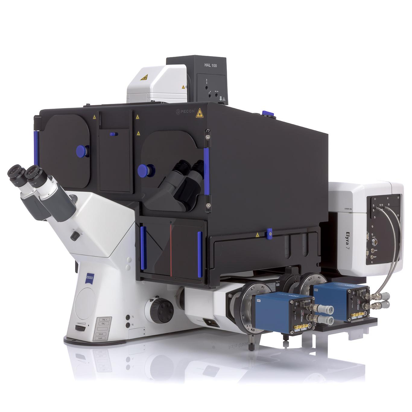

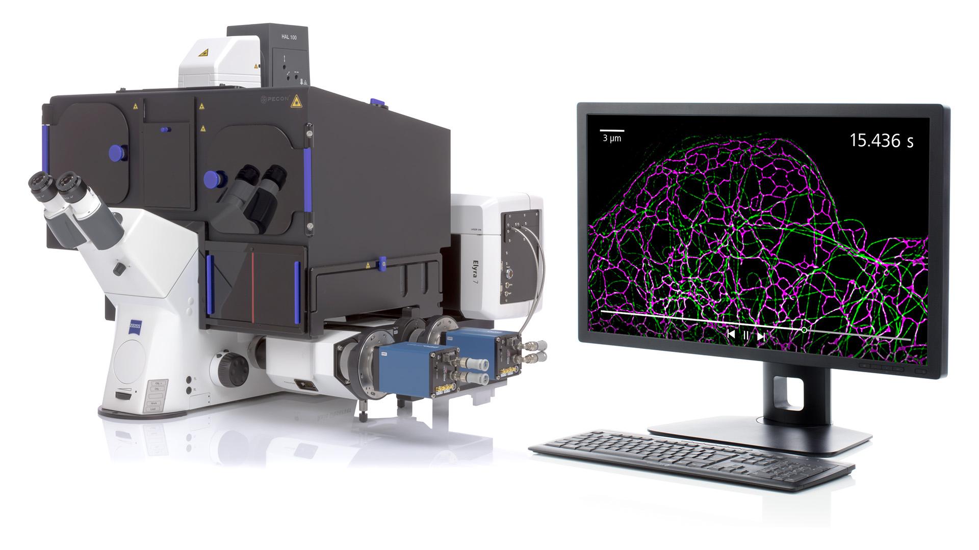
ZEISS Elyra 7 with Lattice SIM²
Your Live Imaging System with Unprecedented Resolution
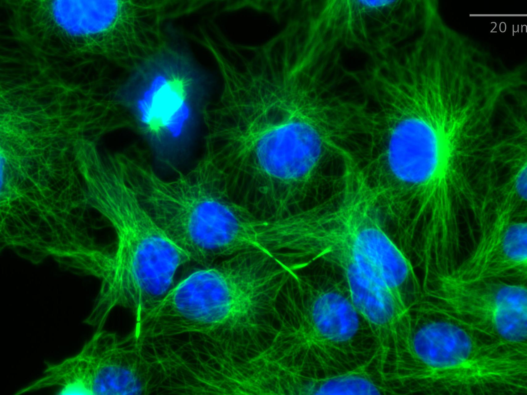
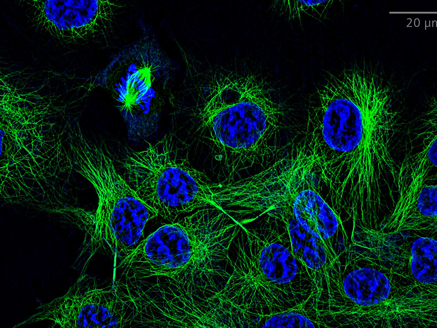
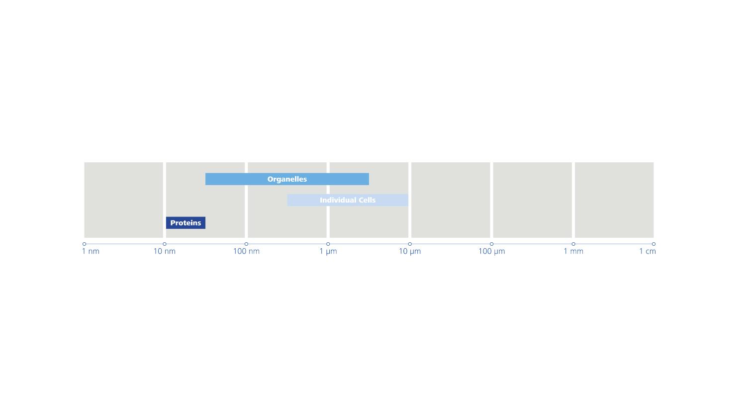
The super-resolution microscope Elyra 7 takes you far beyond the diffraction limit of conventional microscopy: With Lattice SIM² you can now double the conventional SIM resolution and discriminate the finest sub-organelle structures, even those no more than 60 nm apart. You don‘t need to sacrifice resolution when imaging at high speed using only the minimal exposure needed for life observation. Elyra 7 enables you to combine super-resolution and high-dynamic imaging – without the need for special sample preparation or expert knowledge of complex microscopy techniques.

SIM²: Double your SIM Resolution
What if you could push the resolution even further – down to 60 nm?
Lattice SIM opened the door to new applications by enabling gentle super-resolution imaging down to 120 nm at incredible high speed and with the ability to image deeper into challenging samples.
With Lattice SIM² you can now double the conventional SIM resolution.
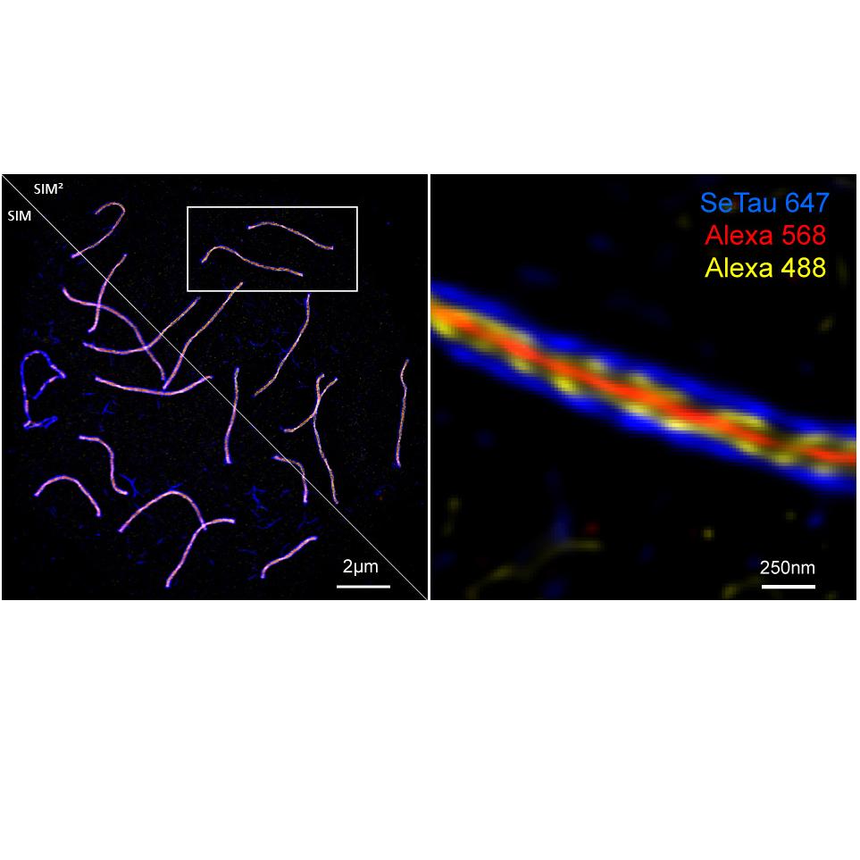
SIM² is a novel image reconstruction algorithm that increases the resolution and sectioning quality of structured illumination microscopy data and raises the SIM technology to a new level. With SIM² you can now discriminate sub-organelle structures down to 60 nm without the need for special sample preparation or expert knowledge of complex techniques.
Watch the video to see the comparison of SIM to SIM² sectioning.
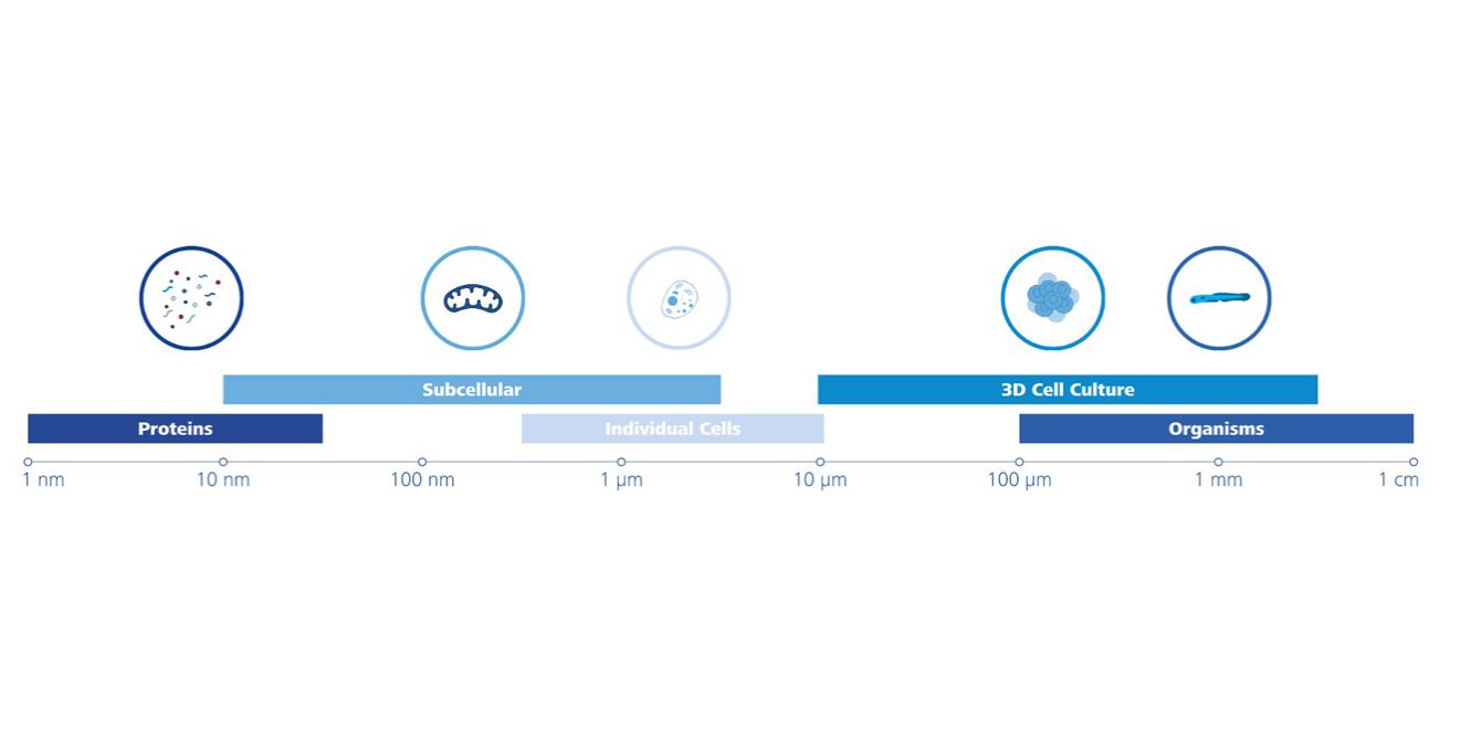
Typical Applications

-
SIM2
- Reveal mechanistic details in live cells, e.g. moving organelles, vesicle trafficking, membrane reorganisation
- Resolve structural details in 3D and multiple colours. Acquire up to four colours with optimized resolution for each wavelength.
- Discover fast cellular processes in the context of whole cells.
SMLM
- Track many molecules over a large FOV and retrieve diffusion behaviour information in entire cells.
- Study molecular level structural changes of sub-minute-scale dynamic processes, e.g. mechanisms of focal adhesions, reorganization of tubulin, vesicle shuttling.

-
- Resolve structural detail in 3D with high penetration depth
- Resolve structural details in 3D over large areas using tiling
- Resolve structural detail in 3D with high penetration depth
-
SIM2
- Probe the structural organization of a whole cell with the advantage of fluorescence specificity and superresolution.
- Investigate arrangement of cellular components and proteins
- Explore interaction of molecules.
SMLM
- Reveal the ultrastructue of organelles and molecular assemblies
- Explore interaction of molecules


From Image to Results
Image analysis? No Problems
Translate your images into quantitative data
ZEISS ZEN
ZEN software provides you with a variety of different image pre- and post-processing tools. For example, you can take advantage of machine learning based on the ZEN Intellesis module to segment complex image data in an easy and intuitive way.
arivis Vision4D®
Use the efficient arivis Vision4D® software forvisualization and quantification of large 3D and 4D data sets imaged with your Elyra 7.
Vision4D® provides advanced image processing tools such as volume fusion, channel shift, conventional and machine learning based segmentation and 3D tracking. Visualize your quantitative results within the arivis Vision4D® software or export all data for further analysis.